FOR MEDICAL INSTITUTIONS

Turn idle MRI capacity into new value for your brain screening.

BrainSuite is examination software that uses AI to measure hippocampal volume from the head MRI (3D T1) images you already acquire. It fits into an existing brain screening programme with no additional scanning burden, and generates a report in about 20 to 30 seconds.

A physician explaining a BrainSuite report next to an MRI scanner

No installation fee / No monthly fee / Usage-based only

Installation fee

¥0

No up-front cost

Monthly fee

¥0

Usage-based only

Analysis time

20–30 sec

Automatic, after the scan

Available since

2021

Japan, China, Hong Kong, Taiwan

ISSUE

Do any of these sound familiar?

01

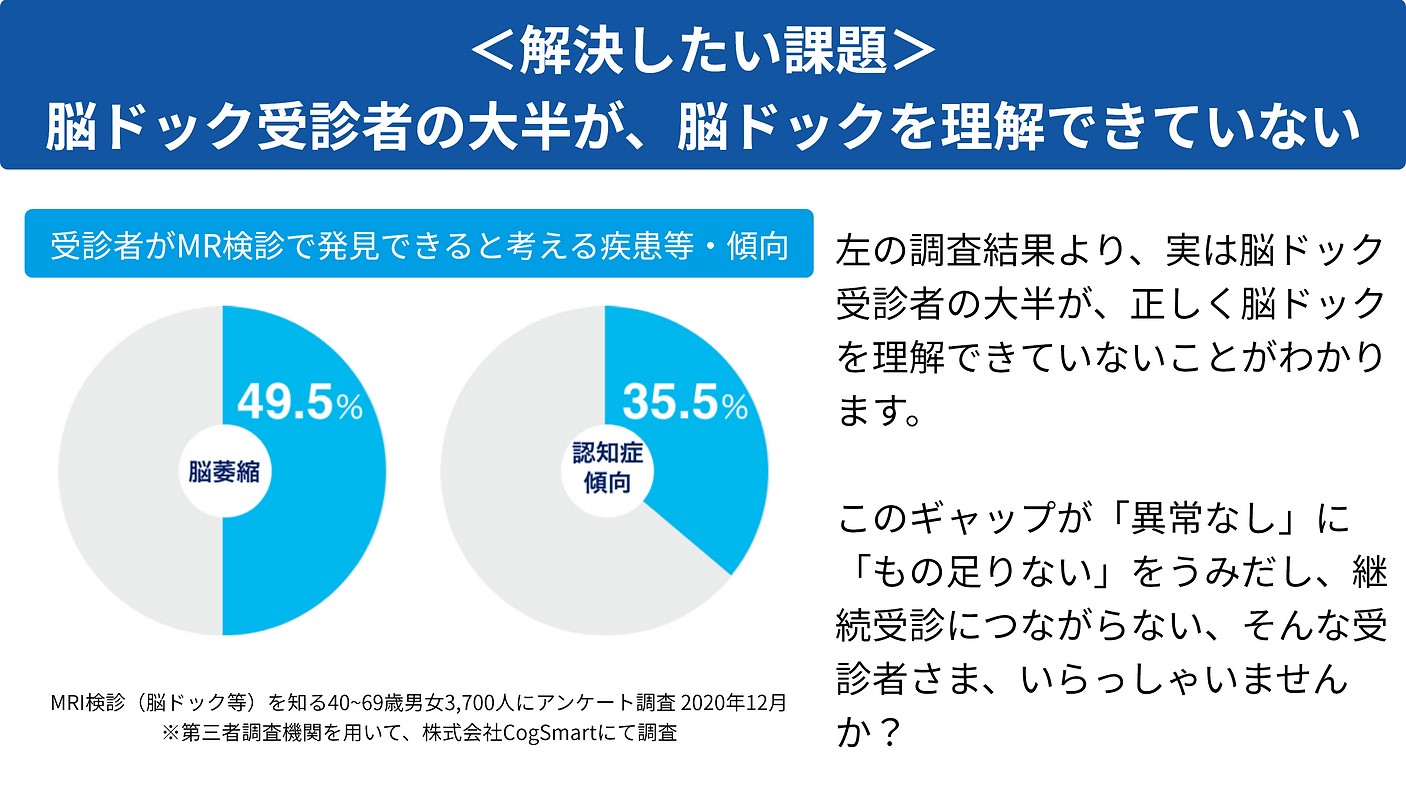
Brain screening ends at “nothing abnormal”

With no findings, the patient leaves with little in hand, which makes it hard to give them a reason to come back.

02

Hard to stand out from nearby providers

With the same scan and the same explanation, price becomes the only reason to be chosen.

03

You want to raise MRI utilisation

There are only so many self-funded examinations you can add, so utilisation and revenue are hard to grow.

04

You want to strengthen follow-up after the examination

Supporting lifestyle change after the results are handed over is hard for a facility to carry alone.

Because BrainSuite can be performed together with the contents of a standard brain screening, it enriches the content and improves the profitability of brain screening — and the rate at which patients return.

WHAT IS BRAINSUITE

AI software for hippocampal MRI examination

Conventional brain screening (MRI/MRA) cannot quantify subtle hippocampal atrophy. BrainSuite analyses and reports it with AI informed by research at the Institute of Development, Aging and Cancer, Tohoku University. Reporting the degree of atrophy while a person is still well supports them in maintaining their brain health.

Analyses the head MRI (3D T1) images you already acquire. No additional sequence and no contrast agent.

Quantifies hippocampal volume in cubic millimetres and reports it against the same sex and age group.

An examination that speaks to brain health while a person is still well, rather than after symptoms appear.

Sample BrainSuite report
▲ The report handed to the patient (hippocampal analysis). Japanese-language sample.

4 REASONS

Four reasons to adopt BrainSuite

AI analysis: automatic extraction of the left and right hippocampus

AI analysis: automatic extraction of the left and right hippocampus

01

Easy to add to brain screening, and it improves MRI profitability

No installation fee and no monthly fee — usage-based pricing only. AI analyses the 3D T1 image and generates a report in about 20 to 30 seconds. It can be added as an option to existing slots, contributing to both MRI utilisation and profitability.

Little or no additional scanning time

Can be offered on its own or as a brain screening option

Each institution sets the price offered to patients

02

It brings patients back

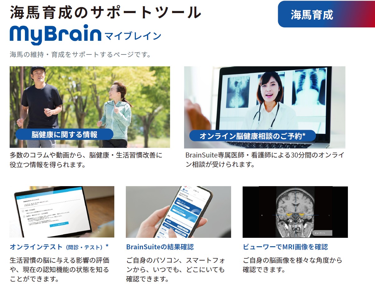
We take on the follow-up after the examination through MyBrain, our hippocampus support tool, and BrainUp, our hippocampus training app. Turning results into preventive action, and making change over time worth measuring, leads to regular repeat visits.

Your team doesn't absorb the follow-up burden

Year-on-year comparison is a reason to return to the same institution

03

Medical evidence and AI image analysis

The software makes the technology and databases used in brain research at the Institute of Development, Aging and Cancer, Tohoku University available for clinical use. Hippodeep, published in international journals, was developed through deep learning on some 25,000 head MRI images.

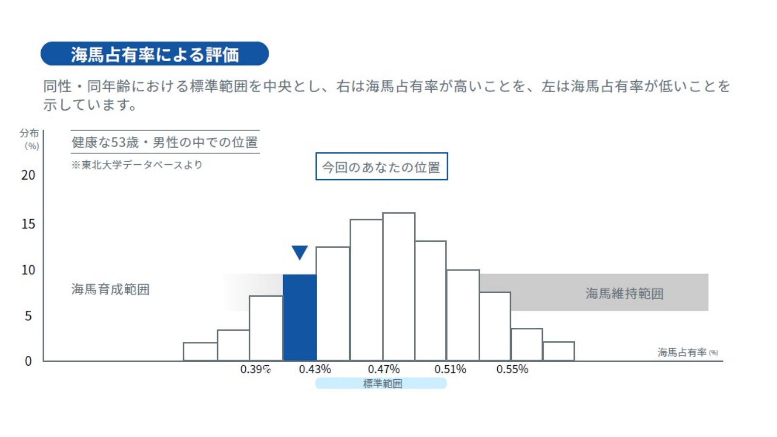
Shows the atrophy trend against the same sex and age group

Includes a simulation of change over the years ahead

04

Support that extends to digital operations

We also provide consulting aimed at patient acquisition through digital transformation and at making booking operations more efficient — supporting hospital and clinic management alongside BrainSuite.

Advice on web acquisition and booking journeys

Patient-facing print materials are available

REQUIREMENTS

All you need is an MRI scanner and a PC with an internet connection.

01

An MRI scanner that can acquire 3D T1 images

We analyse 3D T1 images acquired on the scanner you already use. No dedicated hardware purchase.

02

A PC with an internet connection

Used to upload images and receive reports. No on-site server required.

03

Agreed operating rules

We design how patients are informed and how results are handed over together with you at onboarding.

We are happy to advise on whether your scanner can acquire 3D T1 images and on the acquisition parameters. Send us the model name and we will confirm.

Ask about parameters

WORKFLOW

On the day of the examination

BrainSuite slots into your existing brain screening flow without changing it.

1As usual

Acquire the head MRI

Capture the 3D T1 image within your brain screening protocol.

2A few minutes

Upload the image

Send it from the browser. No dedicated terminal.

3About 30 seconds

AI analyses it

Hippocampal volume is calculated and the report is generated automatically.

4Same day onward

Hand it to the patient

Use the report as it is in the results consultation.

Report page comparing the patient with the same sex and age group over time
▲ Comparison with the same sex and age group, and a projection of change over time.
Head MRI image; the hippocampus is highlighted in yellow

EVIDENCE

Medical evidence and AI image analysis

BrainSuite makes the technology, databases and analytical approach used in brain research at the Institute of Development, Aging and Cancer, Tohoku University available in clinical practice. Hippodeep, the AI image analysis technology behind it, has been published in international journals and was developed through deep learning on some 25,000 head MRI images.

Combined with Tohoku University's brain imaging database, BrainSuite does more than measure hippocampal size: it shows the atrophy trend relative to the same sex and age group, and a simulation of how it may change in the years ahead.

AFTER THE EXAM

Follow-up after the examination

Our services take on the follow-up that comes after the results consultation, so patients are carried through to their next visit without adding to your workload.

MyBrain

Hippocampus support tool

Patients can review their results, track change over time and receive lifestyle guidance themselves.

BrainUp

Hippocampus training app

Daily activity logging connects the examination result to preventive action.

ONLINE

Brain health consultations

Physicians and nurses on our staff respond to patients' questions online.

MyBrain on desktop and smartphone
▲ MyBrain, the patient's personal page

PRICING

Pricing

No installation fee, no monthly fee —
usage-based pricing only

Installation fee

¥0

Monthly fee

¥0

Analysis fee

Per use

Each institution sets its own examination price and the price offered to patients. Request our materials for details.

STEPS

Getting started

1

Enquiry / request materials

Send us your institution's details through the form.

2

Online briefing

We walk through the service and how it would run.

3

Check acquisition parameters

We confirm that your scanner can acquire 3D T1 images.

4

Agreement and operational design

We set pricing and how patients will be informed.

5

Go live

After a test analysis, you begin accepting patients.

Frequently asked questions

Q. Do we need dedicated hardware or a server?

A. No. An MRI scanner able to acquire 3D T1 images and a PC with an internet connection are enough.

Q. How much longer does scanning take?

A. If you already acquire 3D T1, there is normally no additional acquisition time. Parameters can be discussed case by case.

Q. Does it add to reading or consultation workload?

A. Reports are generated automatically and can be used as they are in the results consultation. We take on the follow-up afterwards.

Q. Is the price to patients fixed?

A. Each institution sets it. We are happy to advise with the local situation in mind.

Q. Can it be offered as a standalone examination?

A. Yes. Institutions run it both as a brain screening option and as a standalone examination.

Start with the materials, and see how BrainSuite fits your programme.

We will send a pack covering the service, a sample report, the operational flow and the pricing structure. Online briefings are also available.

Request materials (free)

Institution name, your name, email and phone number only. It takes about a minute.

A physician explaining results to a patient

About CogSmart, Inc.

A medical and healthcare company spun out of the Institute of Development, Aging and Cancer, Tohoku University

Head office

Room 604, Nihonbashi Life Science Building 2, 3-11-5 Nihonbashi-honcho, Chuo-ku, Tokyo 103-0023, Japan

Representative Director

Akira Higuchi

Science and Technology Advisor

Yasuyuki Taki

Business

BrainSuite®, AI-based hippocampal MRI examination / BrainUp®, an app for training the hippocampus / data science grounded in neuroscience and medical evidence

Dr. Yasuyuki Taki

SCIENCE & TECHNOLOGY ADVISOR

Yasuyuki Taki (MD, PhD)

Professor, Department of Clinical Research on Aging, Institute of Development, Aging and Cancer, Tohoku University; Director, Smart Aging Research Center
Science and Technology Advisor, CogSmart, Inc.

Brain health is not fixed at birth. It can be changed considerably by what we do each day. The hippocampus, which governs memory and emotion, is a particularly important region: exercise, learning and good daily habits can help it grow.

BrainSuite is a tool not only for knowing the state of the hippocampus, but for using that knowledge as a reason to change behaviour. My hope is that people take a first step for their hippocampus today, for the sake of their future selves.